Monday, June 30, 2014

“Liquid Biopsy” – measuring cell-free circulating donor DNA to predict allograft rejection

Transplant physicians face the challenge to detect allograft rejection using non-sensitive biomarkers and clinical signs/symptoms. 
For kidney transplant, serum creatinine, urinalysis for proteinuria or hematuria, are so far used for screening for rejection and/or assessment of graft function. In a recent article in Sci Transl Med, De Vlaminck et al. investigated the use of cell-free circulating DNA (cfdDNA) as a marker of allograft rejection in adult heart transplants. 
Authors used shotgun sequence approach to detect donor-specific DNA fragments (SNP genotyping), and correlated the level of cfdDNA with cardiac biopsy results, the diagnostic “gold standard” for rejection. 
The idea of a “liquid biopsy” —using plasma circulating DNA fragments—as a diagnostic tool has been explored in the Oncology field, to detect early and late stage cancers. In transplant field, the presence of donor-derived DNA fragments in recipient’s plasma (or urine), called microchimerism, has been known since the end of 1990s  and using them as a measure of transplant injury has been tried. For example, Sidgel et al. used Y-specific DNA fragments as a marker in male-to-female transplants. 
 In this current article, De Vlaminck et al. further improved this approach to overcome dependency on donor-recipient pair. Authors concluded that using the cutoff value of 0.25%, cfdDNA method is as sensitive (0.58) and specific (0.87) as biopsy (0.58 and 0.96, respectively). Furthermore, cfdDNA approach was able to detect rejection as early as 5 months prior to tissue diagnosis
There are, of course, some limitations to overcome: 1) cfdDNA cannot differentiate ACR vs AMR, 2) cannot rule out other source of DNA fragments, such as endothelial damage without rejection 3) exhibit still substantial discordance with biopsy results. Although currently using the shotgun approach, the technique can be refined by using targeted-sequencing approach, to be less laborious process. 
Based on the same underlying principles, it is very likely that similar test would have application in kidney transplantation and potentially help to detect earlier ongoing graft injury. We will still need the biopsy though to clarify the type of injury...

Naoka Murakami

Monday, June 23, 2014

IgM Nephropathy: What You Need to Know


Similar to my recent post on C1q Nephropathy, IgM Nephropathy (IgMN) is a podocytopathy that is often considered a variant of Minimal Change disease (MCD) or FSGS. Like C1QN, it is also a controversial entity with a lack of uniformity from a diagnostic point of view. This undoubtedly leads to under-reporting with some pathologists calling a case IgMN what others would call MCD/FSGS/Mesangial Proflierative GN with low level IgM staining.
First descriptions of IgMN reported mesangial hypercellularity on light microscopy, granular IgM and C3 mesangial deposits on immunofluorescence with electron dense deposits in about half of cases. Podocyte foot process effacement was also evident consistent with the presentation of nephrotic syndrome. The incidence has been reported to be 2-18% of renal biopsy series and age at presentation appears to be bimodal with a peak in childhood and again later in life, 6-7th decades. The incidence of IgMN may be higher in the developing world and most recent reports come from developing nations (ref, ref).
The accurate diagnosis of IgMN is important from a prognostic and therapeutic point of view. Overall, IgMN does not respond to steroids as well as MCD with a higher proportion of patient with steroid-dependent and resistant disease. Furthermore, assessment of children with steroid-dependent and resistant MCD revealed a high proportion of IgMN, which appeared to respond better to cyclosporin than to cyclophosphamide. Rituximab (as ever!) may be another alternative therapeutic choice, although evidence remains at case report level, including a case of recurrent IgMN post-transplant. Similar to C1QN, the severity is likely dependent on the light microscopic changes with MCD patterns reporting an excellent remission rate and an FSGS pattern of injury having a worse prognosis (ref). Most cases however may have mesangial proliferative changes evident. There is even a report of IgMN presenting with crescentic lesions.

IgM deposition may be seen in many other glomerular disorders and its role is unclear. Is it an innocent bystander, reflecting natural IgM binding to exposed epitopes on injured glomerular cells? There is some evidence that IgM deposition may activate complement and that efforts to prevent IgM antibody deposition may prevent complement activation and slow the progression of glomerular injury. IgMN is another glomerular disease that we have an unclear understanding of pathogenesis and etiology where we rely on crude pathological descriptions. Hopefully the future will bring clarity to these diseases and facilitate precise molecular diagnosis of entities such as IgMN.

Sunday, June 22, 2014

POSEIDON Trial for Contrast Induced Nephropathy: The Next #NephJC

The next Nephrology Twitter Journal Club (@NephJC) will discuss the POSEIDON Trial which has been published in The Lancet. Interest in the prevention of Contrast Induced Nephropathy (CIN) is alive and well with several novel approaches/therapies being recently reported (see my recent post), mostly in non-Nephrology journals. Despite these interventions, volume expansion remains the cornerstone of preventative management for CIN (contrast-induced acute kidney injury is just too much of a mouthful for me).

This current trial randomized patients into 2 protocols of 0.9% NaCl-based volume expansion peri-cardiac catheterization. The study group had their fluid rate governed by left ventricular end diastolic volume, measured easily during the catheterization procedure and used as a surrogate for volume status. This allowed the intervention group patients to safely receive higher volumes of saline during their procedures which translated into significantly lower rates of CIN. Obviously this method can only be conveniently employed during catheterization procedures and would need the involvement of interested cardiologists for it to be used in real-life clinical practice. However, the results appear impressive.
You can read a concise summary of the trial and results over at NephJC. Be sure to tune into Twitter on Wednesday June 25th at 9pm Eastern and feel free to get involved using #NephJC.

Monday, June 16, 2014

Exams Are Coming

This season's Game of Thrones finished last night and as a nephrology-themed homage, a trainee from Malaysia, Lee Jun, created this poster during downtime while studying for his exams. Enjoy.

Saturday, June 14, 2014

C1q Nephropathy: What You Need to Know

I saw a 20 year old African American male in clinic with nephrotic range proteinuria, reduced GFR and no signs or symptoms of a systemic disease. His mother developed ESRD at age 36 and his brother, aged 26, has CKD Stage 4. A full laboratory workup including complement proteins and ANA was unremarkable. Renal biopsy demonstrated an FSGS pattern of injury with a full house picture on immunofluorescence but with dominant C1q staining. A diagnosis of C1q Nephropathy (C1QN) was made. 

Jennette et al described C1QN in 1985 in 15 patients without evidence of lupus who had dominant mesangial C1q deposition, often with C3 and Ig’s also. The mean age at presentation was almost 18 years, with a similar male: female ratio and average proteinuria of 7.5g/day. Another series from Columbia University included 19 patients who were predominantly African American (73%), young and female (73%) with nephrotic range proteinuria. Renal outcomes across the series’ (including this European series) are mixed but would suggest immunosuppressive treatment directed against the primary pathology should be considered.
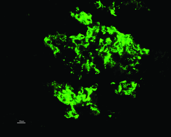
C1QN may be considered an independent pathological entity, although this has been controversial, with some classing it as a subtype of FSGS or minimal change disease (MCD). The prevalence varies (0.2-16%) but is more common in children. The predominant light microscopic appearance may be MCD, FSGS or a proliferative glomerulonephritis. While C1q deposition (predominantly in the mesangium; see image) is dominant or co-dominant by definition, a full house pattern of IgM, IgG, IgA, C1q and C3 may be observed, more commonly with a proliferative glomerulonephritis. As for treatment, the case series are small and largely uninformative. Some patients appear to remit with or without immunosuppressive treatment whereas others have a rapid course towards ESRD. Patients with an FSGS pattern may do worse than other types. For what it’s worth, my patient had no response to 6 months of steroids and mycophenolate and a plan for cyclosporine was made. The fact he likely has a genetic element to his disease adds complexity. Some would argue that immunosuppressive therapy in these cases is not likely to be of benefit but we see from our hereditary FSGS cohort that this is not necessarily the case. Cyclosporine and Rituximab in particular appear to have a directly beneficial effect on podocyte function (also see case report of Rituximab treatment in C1QN).
As for the pathogenesis and significance of the C1q deposits, we have much to learn. Are the C1q deposits pathogenic or merely markers of injury? We do know that C1q binds to the basement membrane via laminin and C1q receptors enhance binding of immune complexes to mesangial cells where it combines with other complement proteins to form C1 protease. This in theory allows activation of the classical complement cascade. Despite this, serum C4 levels are generally normal and I can find no reports of complement directed therapy (Eculizumab or C1-Inhibitors) in C1QN.
Bottom line: C1QN is likely an under recognized entity which can have varying clinical and histological presentations. Renal outcomes are also variable and it may have a different prognosis compared to traditional MCD (& FSGS?). One to keep in mind for the boards.

Friday, June 13, 2014

Immortality

A few months ago, a paper was published in the International Journal of Epidemiology that caused a sensation in it's home country - Denmark. Using registry data, and the diagnosis of non-melanomatous skin cancer as a proxy for sun exposure, the authors found that the OR for mortality in individuals who developed skin cancer compared to those who did not was 0.53. The p-value was an unfeasibly low 2x10E-308. However, when the results were stratified by age, the OR was a much more reasonable 0.97 in the sun-exposed group. This fact was conveniently forgotten when the study was reported in the media which uncritically stated that people who get more sun will live longer - something which upset the Danish cancer societies immensely.

So why was there such a disparity between the two results and how could it be possible that individuals with cancer could live so much longer (8.5 years on average) than those who did not get cancer. This month, a mea culpa editorial was published in IJE explaining their mistake along with an article discussing the entire issue. What the authors did in this case was fail to account for the immortal time bias. In this study, individuals entered the cohort when they were 40 years old. However, most people did not develop skin cancer until they were in their 60s or 70s. As a result, for the skin cancer cohort, there were approximately 20 years where they could not have died (they had to survive until they developed cancer and were "immortal" until that time). Someone who did not develop skin cancer could have died at any time during that 20 year period. To demonstrate how this works, the authors of the follow-up study, using the same dataset, randomly allocated a "lottery prize" to a proportion of individuals with a mean age of 68 who lived in Denmark over the last 20 years. Again, in this case, a person who received the prize would have had to have lived until the time that it was awarded while those who did not get the prize could die at any time. The results of the simulation study were very similar to the skin cancer study with an OR for all-cause mortality of 0.5 for the prizewinners and a similarly outrageous p-value.

This issue was first described in the 1800s when it was noticed that generals and bishops live longer than lieutenants and curates. Again, this is because one has to survive to an older age to become a bishop or a general and not because there is something inherent in these ranks that lead to an improvement in mortality.

This example is particularly egregious and the editors of IJE have to be congratulated for the way that they dealt with it. However, there are more subtle examples, one of which, highlighted in the follow-up article, appeared in JASN in 2010. This paper found that survival after transplant failure was improved by nephrectomy. However, the mean follow-up in this paper was only 2.93 years and the mean time to nephrectomy was 1.66 years. Thus, about half of the follow-up in individuals who had a nephrectomy was immortal time - they could not die during that follow-up period because they had to survive to the time of surgery. Most of the difference could be accounted for by this bias.

Similarly, a study in JASN in 2007, found that individuals enrolled in a multidisciplinary care clinic were more likely to survive than those who were not. Again, the time of the first MDC clinic was about 1 year after enrollment in the study. Individuals who entered the MDC program had a year of immortal time compared to individuals who did not enter MDC. This was pointed out in a follow-up article in KI. The authors of the original paper reanalyzed their data to account for this bias and found that MDC was still associated with improved survival although the magnitude of the effect was substantially less.

This is a fascinating issue and probably affects more cohort studies than we think. As a reviewer, I'll certainly try to look out for it in the future.

Wednesday, June 11, 2014

Unusual transplant immunology case

File this under something that we are never likely to see in clinical practice. In 2002 a fascinating case report was published in the New England Journal of Medicine concerning unusual findings during a work-up for a living related kidney transplant. A 52-year old woman had FSGS and was approaching ESRD. Her three sons were willing donors but when they went for HLA typing, there was a problem. Although all of her sons had a HLA haplotype from their father, two of the three had a HLA haplotype which, while identical to each other, did not match either of their mother's HLA haplotypes suggesting that she could not be their mother! Further investigation revealed that her sons' non-paternal HLA haplotype was identical to that of the patient's brother and had ultimately been derived from her deceased father.

Further analysis of the patient's blood found that there was no evidence of two distinct cell populations. However, hair follicles, buccal mucosa, bladder samples from a prior cystoscopy and thyroid cells from a prior resection found 4 haplotypes in these tissues. Only two were present in her peripheral blood. There was a normal number of chromosomes in all cells. This was a case of tetragametic chimerism. It is likely that there were two fertilized embryos in utero and that they fused at some point during development to form a single individual. It turns out that under experimental conditions inducing chimerism in mice, it is not unusual for such individuals to express a single haplotype pair in the peripheral blood while continuing to have all 4 haplotypes in other tissues. No-one really knows how common this condition is although the authors suggest that it may be more common than we appreciate. It has important implications in particular for forensic science.
One of the more interesting aspects of this case was that the patient had a HLA identical brother and a HLA haploidentical brother. Although she appropriately lysed cells from control individuals, she did not react to cells from her brother despite the fact that her lymphocytes carried a different HLA haplotype. She was fully tolerant to her brother.

A couple of years later, a woman in Washington State was in the middle of a paternity suit after separating from the father of her two children. She was pregnant with the third child at the time. Testing revealed that he was indeed the father of the children but that she was not. She was charged with fraud and accused of taking part in a surrogacy scam. Her children were taken off her and when her third child was born, this child was tested too and also found not to be her child. Her lawyer found this case report and asked for further testing - results of a smear test revealed that the DNA in that sample matched her children - she was a chimera also. Her children were promptly returned.

Monday, June 9, 2014

Hyponatremia Guidelines - NephJC

Tomorrow night will be the 4th edition of the Nephrology Journal Club on twitter.The article this week is unusual - it is a clinical practice guideline for the management of hyponatremia that was published in NDT, the European Journal of Endocrinology and Intensive Care Medicine. It is an interesting piece of work that has been expertly summarized by Joel Topf over at PBFluids. Even if you do not feel up to reading the entire paper, consider having a look at his executive summary as it raises a number of important (and eminently debateable) points.

See also the summaries at NephJC.com and Nephron Power. Join everyone for what is likely to be a lively discussion tomorrow at 9pm ET on twitter using the hashtag #NephJC.